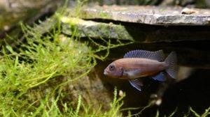
Aquarium keeping is a fulfilling and rewarding hobby, but when our fish show signs of illness, it becomes an immediate source of stress for us. Here’s the good news: If your fish is struggling with swim bladder disease, its symptoms are fairly easy to recognize.
If your fish is bloated and having buoyancy issues, and you suspect they might have a problem with their swim bladder, keep reading to find out what might be going on and how you can help.
In This Guide
What Is Swim Bladder Disease in Fish?
To understand swim bladder (aka air bladder) disease, you must first understand the swim bladder itself. The primary function of the fish swim bladder is to maintain buoyancy, though it can also play a role in your fish’s hearing and vocalizing. In general, fish regulate the amount of air in their swim bladder in one of two ways: by gulping air at the surface (e.g., carp, koi, catfish, goldfish) or through gas exchange via an intricate network of blood vessels (e.g., cichlids, perch-like fish). The former is categorized as physostomous, and the latter as physoclistous.
When a fish has trouble regulating their swim bladder, it’s commonly called swim bladder disease. Note that swim bladder disease is not a specific sickness but rather a syndrome; it is a sign of other underlying issues. The actual swim bladder is often not the source of the disease but is the organ impacted by the primary illness. More accurately, swim bladder disease can be described as a buoyancy disorder and can manifest positively (floating to the surface) or negatively (sinking to the bottom).
Why Is My Fish Swimming Upside Down?
Sometimes the swim bladder becomes so over-inflated that it is impossible for the fish to counteract the positive buoyancy of the bladder with their swimming movements, resulting in your fish swimming upside down like they’re attached to a balloon.
What Causes Swim Bladder Disease in Fish?
A wide variety of issues can cause the buoyancy problems associated with swim bladder disease, including:
- Poor water quality
- Overeating/improper diet
- Sudden temperature swings
- Bacterial infections
- Parasites
- Other impaired organs
- Physical trauma from shipping, improper handling and fighting with other fish
Because there are so many issues that can present as a buoyancy disorder, it can be difficult to correctly diagnose and treat the underlying cause of swim bladder disease.
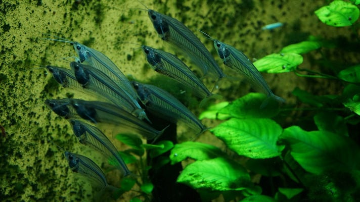
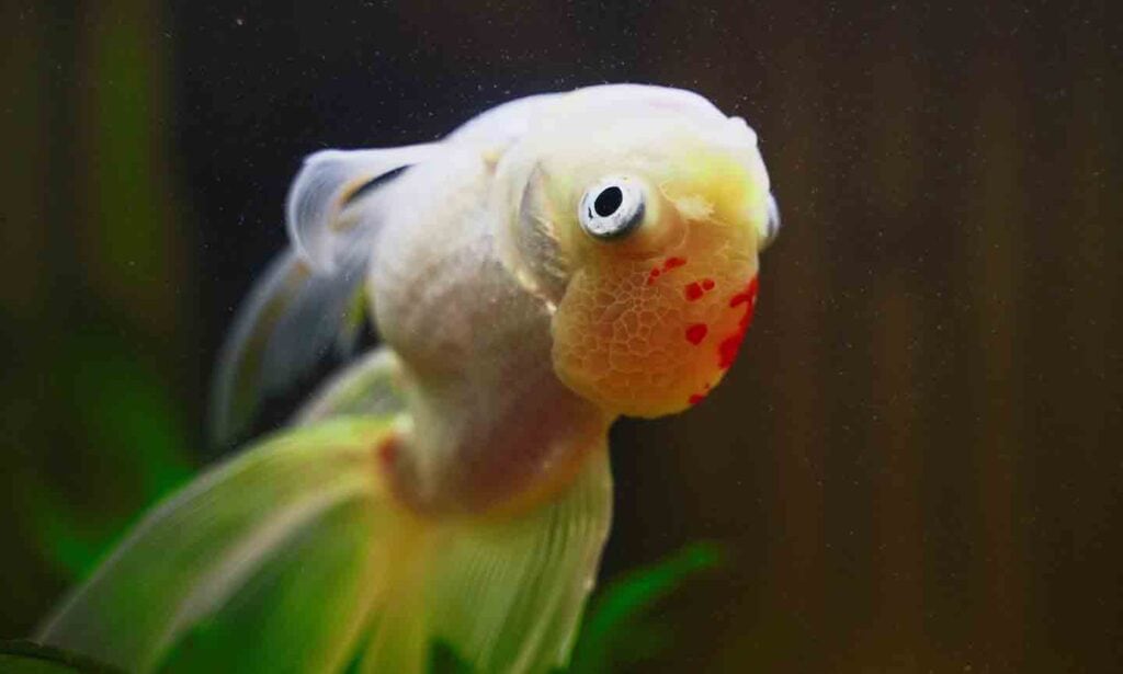
Swim Bladder Disease Symptoms
All symptoms of swim bladder disease involve swimming problems relating to neutral buoyancy, and your sick fish can exhibit one of more of these at once.
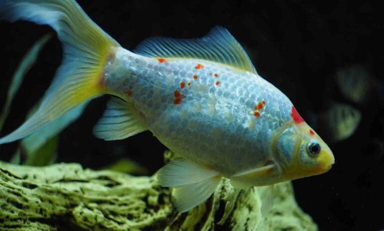
- Distended belly: Your fish’s abdomen will appear full or bloated and the fish will struggle to maneuver normally.
- Floating on the surface: Your fish spends most of their time floating at the surface, potentially upside down in more advanced cases, and has a difficult time descending in the water column, if they can do so at all.
- Sinking to the bottom: Your fish spends most of their time lying on the bottom of the tank and struggles to swim to the surface, if they can do so at all.
- Listing, leaning or swimming on one side: Your fish cannot stay upright, and they always seem to be tipping to one side, making it difficult or impossible to swim normally.
- Head tilted up or down: Your fish’s head is either tilted upwards or downwards, and they cannot seem to right themselves even with great effort.
Being constantly exposed to air or regularly resting on the substrate can cause additional medical issues because these behaviors have a negative impact on the slime coat, and breaking down this natural barrier makes fish susceptible to a host of infections. Struggling to swim normally is also extremely stressful and energy intensive, further contributing to a decline in their health.
Diagnosing Swim Bladder Disease in Fish
To unequivocally diagnose a fish with this syndrome, the fish would need to be taken to a veterinarian for X-ray examination and a prescribed treatment regimen. As this is an unlikely course of action for the vast majority of home aquarists, we must do our best with the simple information we have. If your fish’s body is bloated and they’re having trouble regulating their buoyancy in any of the ways mentioned in the section above, there is a good chance there is something wrong with their swim bladder.
Swim Bladder Disease Treatment
We can’t treat anything effectively unless we know what caused the issue in the first place, and because pinpointing the cause of swim bladder disease is nearly impossible to do without advanced testing methods, you may need to make some educated guesses. Follow these steps to confirm or rule out the most likely causes:
1Consider Your Water Quality
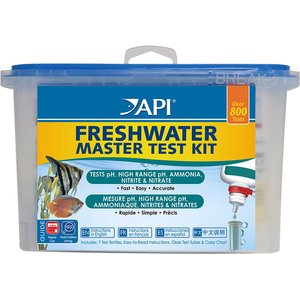
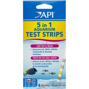
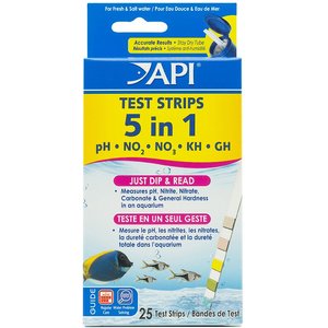
The most overlooked contributor to swim bladder disorders is poor water quality. If your fish is having a buoyancy problem, the first thing you should do is measure some water quality parameters, including:
- Ammonia level
- Nitrite level
- Nitrate level
- pH of the water
All common aquarium test strips will be able to measure these basic water quality parameters.

Any level of ammonia or nitrite
Substantial water changes (25-50% of the tank) until levels are no longer detectable
High nitrate levels
Substantial water changes (25-50% of the tank) until the level returns to an acceptable rate for your fish
pH too low or too high
Use an aquarium water treatment designed to raise or lower your tank’s pH
The recommended range for all of the above will vary depending on the type of fish you have, so consult a vet or other fish expert about the ideal levels for your tank.
2Consider Your Water Temperature
Temperature stress can also lead to swim bladder disease. A water temperature that shifts rapidly (such as from a malfunctioning heater or chiller) can absolutely cause swim bladder disorders, as can temperatures that are chronically too high or too low. If, for example, you’re keeping a tropical fish species that prefers water temps in the low 80s, but the water is consistently in the low 70s, this low water temperature will chronically stress your fish, lowering their immune system and making opportunistic infections more likely.
Talk to your vet or fish expert to find out what the ideal temperature is for your fish species, and use a thermometer to monitor the temperature of your tank. You can use a heater or chiller to bring the temperature higher or lower. If your temperature is unstable, it may be time to repair or replace your heater or chiller.


3Consider Your Fish’s Diet
Improper diet is another major factor that causes swim bladder disorders. Feeding the wrong foods or too much food can lead to gut distension and digestive tract blockages that obstruct the pneumatic duct (a structure that connects the swim bladder to the digestive tract in some fish), leading to swim bladder disease.
First, ensure that you’re feeding your fish the correct food for their species. Many types of fish food list their appropriate fish types on the packaging and/or in the product description.
Secondly, watch your fish during and after their meals. Large amounts of dried, floating fish foods can expand in the gut and lead to swim bladder disease, so if you notice that your fish are leaving flakes behind to snack on later, you may need to cut down on the amount of food you feed.
If overfeeding is the culprit, it is recommended to fast your fish for two to three days and then slowly start feeding again with high-fiber food items. A poor diet lacking in fiber can cause gas in the gastrointestinal tract, leading to severe constipation that makes the abdomen swell, preventing the swim bladder from functioning properly. Understanding the dietary needs of your fish is crucial to their well-being and a key to successful fishkeeping.
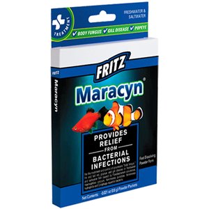
4Try an Antibiotic
Since secondary bacterial infections are one of the most common causes of swim bladder disorders, pharmaceutical intervention may be needed to help your fish. If you take the actions mentioned above and your fish’s condition does not improve, it may be wise to try a broad-spectrum antibiotic, such as Maracyn from Fritz.

Temporarily out of stock
These are just some of the reasons that your fish can have a buoyancy disorder, and this is not meant to be a comprehensive list of every single possible explanation for your fish’s illness. Consult an aquatic veterinarian or other fish expert if your fish is still struggling with buoyancy issues.
Swim Bladder Disease Prognosis
As with many diseases, the sooner you catch this the better. If remedied soon after the onset of buoyancy issues, the fish has a good chance of returning to normal. However, if left untreated for an extended period, the likelihood of recovery and survival is much lower.
Preventing Swim Bladder Disease in Fish
The single best preventative measure for swim bladder problems in aquarium fish is good water quality. Appropriate temperature and pH along with low nitrates and no ammonia or nitrites will go a long way towards avoiding swim bladder disease. Proper diet is also critical, as well as correct tank size and suitable tank mates.
Stress of all kinds leaves our fish susceptible to opportunistic infections that affect the swim bladder, so reducing the overall stress levels by providing your fish with an ideal aquarium setup is a sure path to success. Find out more about alleviating fish stress.
Expert input for this story provided by Dr. Jörg Mayer, DVM, Professor of Zoological Medicine at University of Georgia College of Veterinary Medicine in Athens, Georgia.
Keeping Your Fish Healthy
Share: