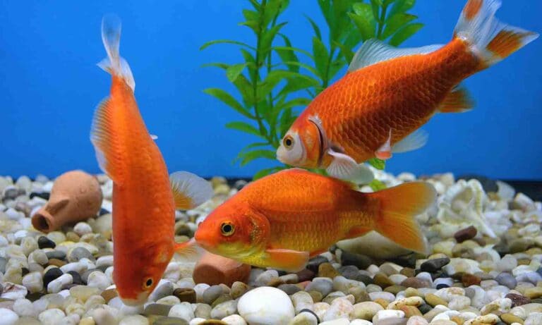
Of the fungus diseases that may affect freshwater fish, body fungus is the most common. It is also one of the tropical fish diseases that some aquarists regularly encounter. As we will see, this need not be the case, because normally this disease is easily preventable.
The usual causes of this malady are fungi of the family Saprolegniaceae, most commonly members of the genus Saprolegnia and the related genus Achyla. In particular, two species most frequently isolated from tropical fish are Saprolegnia parasitica and S. declina; hence, this disease is sometimes referred to as saprolegniosis.
The living fungus is made up of a mass of threadlike filaments, or hyphae, which grow to form mats (mycelium). Some of the hyphae grow downward like roots into the organic matter they are attacking. Other filaments grow outward, producing the fuzzy growths typical of this disease. Eventually, club-shaped, spore-containing sporangia form at the tips of these filaments. When these sporangia are mature, they rupture, releasing masses of spores into the water.
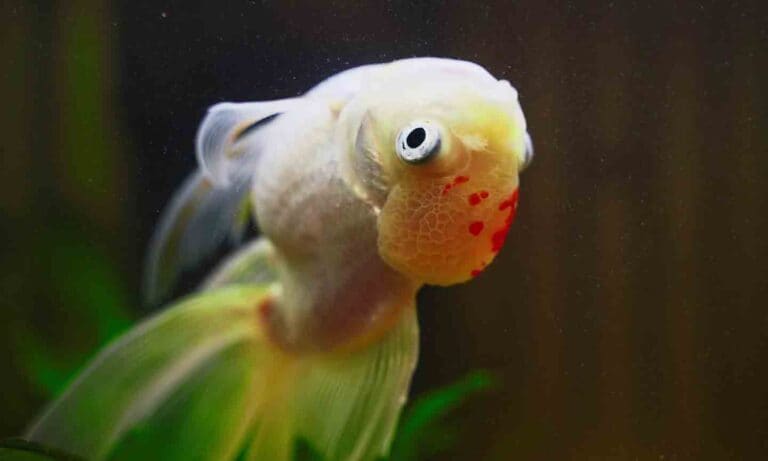
Body fungus is distinctive in appearance and not confused with any other malady. A tropical fish with fungus appears to have one or more slowly enlarging fuzzy patches resembling cotton wool or bread mold (also a fungus) on its body or fins. They are usually white, but minute particles of debris or suspended algae trapped among the fungal hairs can impart a pale tan or greenish color to these growths. The only other diseases that even remotely resemble it are the white lesions of advanced mouth and body rot caused by the bacteria Flexibacter, but these lack the fuzzy appearance. This confusion is perpetuated by the frequent use of the name mouth “fungus” to describe this bacterial disease.
Normally Harmless Inhabitants
Under normal conditions, Saprolegnia fungi are harmless inhabitants of freshwater fish aquariums that attack dead and decaying organic matter. Their spores can be found in just about all aquariums, where they float around in the water until they happen to land on a material suitable for their germination and growth. Common targets are uneaten fish food and dead fish that have not been removed from the aquarium, as well as infertile fish eggs.
All too often, we discover these fungal growths on our tropical fish. With rare exceptions, these outbreaks are a sign of poor aquarium husbandry that could be prevented. Saprolegnia colonizes dead and decaying organic matter, not healthy living tissue – it will not initially attack uninjured healthy tropical fish with intact slime coats, but will secondarily invade the damaged tissues of wounds and injuries. These injuries can be the result of aggression from other tropical fish, rough netting and handling, and tissue damage from external bacterial and parasitic infections. Other common causes are superficial burns from extremes of pH or excessive ammonia build-up.
Ammonia damage to a fish’s skin, gills and fins is frequently seen in newly set-up aquariums with uncycled biological filtration, in overcrowded aquariums and as a result of poor shipping practices in which too many tropical fish are packed in a bag. All these factors also increase a fish’s stress level, weakening its immune system.
Once body fungus gains a foothold on a tropical fish, its hyphae spread rapidly to healthy tissue. Typically, the invasion goes no deeper than the superficial muscle and gill layers, but this damage results in a major loss of tissue fluids and electrolytes. It is also thought that the fungus secretes harmful chemicals that further weaken the immune system. An untreated fish will die in a matter of days from tissue destruction and fluid loss, or suffocation when the gills are involved.
Treatment
When fungus outbreaks appear in your aquarium, you must strive to eliminate the underlying cause. This means eliminating sources of potential or actual injury to the fish, which might take a bit of detective work and involve removing an aggressive tropical fish or sharp rock, checking that the pH and other chemical water parameters are within the desired range, making certain that your aquarium is not overcrowded, and eliminating bacterial or parasitic infections causing injury to the skin and fins. You might also need to improve your fish-netting techniques.
In my experience, body and gill flukes of the genera Dactylogyrus and Gyrodactylus are a major predisposing cause of body fungus infections. These flukes are too small to see without a low-power microscope, but can cause immense, unnoticed damage to the gills and tissues of heavily parasitized fish. Goldfish and discus are particularly prone to fluke infestations.
Complicating the fluke problem is that many strains are resistant to dylox (also known as masotin or trichlorofon), the most common ingredient in typical over-the-counter fluke remedies. I personally recommend that aquarists search out the drug praziquantel, also known as droncit or Prazipro, as treatment.
Attempting to eliminate the fungal spores from an aquarium is not only unnecessary but also impossible. This is because they are resistant to treatment, and are just about everywhere in the air and water.
It is essential to begin treatment at the first signs of disease. Because body fungus is not – in the true sense – a contagious disease, it is best to treat affected fish in a separate container. Tropical fish with large areas of tissue covered by fungus are nearly impossible to successfully treat.
There are many medications sold for the treatment of fungus in fish, such as malachite green zinc-free oxalate (used according to the manufacturer’s instructions – which is important for all medications). This drug should not be used on tropical fish known to be sensitive to this chemical, such as some characoids, many scaleless fish and sunfish of the family Centrarchidae. Note that malachite green/formalin products so successful for ich treatment do not have the malachite green concentration adequate to treat fungus, and formalin should not be used where large wounds are present.
Try Salt
We were once taught that fungi are plants that lack chlorophyll. We now know that this is incorrect, and they are separate from both plants and animals. One thing fungi do have in common with most true plants is their intolerance of salt. This is the reason that body fungus infections are not seen on saltwater fish. Adding 1 tablespoon of noniodized rock salt to each gallon of water is helpful in effecting a cure. In fact, salt might be all that is needed in early cases.
Salt can also be used in combination with malachite green. Salt has another beneficial function: By reducing the osmotic differential between the fish’s body fluids and the aquarium water, it slows the rate of body fluid loss from open lesions. Salt will kill aquarium plants and snails, but this should not be a problem if you are treating the infected fish in a separate container. Remember: You are not trying to eliminate fungal organisms from your aquarium, only from your fish.
As with malachite green, a few freshwater fish, such as Corydoras catfish, are sensitive to salt. Others will require an adjustment of the salt dosage. For example, goldfish and most poeciliid livebearers (such as guppies) can easily tolerate high levels of salt, whereas I would be much more conservative when treating dwarf cichlids and many soft water characins (includes tetras). When using either malachite green and/or salt, treatment should be continued until a few days after a visible cure is achieved.
For Stubborn Fungus
Stubborn cases of body fungus can sometimes be successfully treated with potassium permanganate. There are proprietary formulations of this drug available in pet stores. Follow the manufacturer’s instructions carefully, as it is a powerful oxidizer – it can cause severe and even fatal chemical burns to a fish’s skin and gill epithelium. Potassium permanganate can also become toxic under conditions of high pH, depositing manganese dioxide in tropical fish’s gills. Do not use it with formalin.
Potassium permanganate will tint the water a deep violet, which will slowly fade, and is deadly to plants and biological filtration. Treatment should therefore be done in a separate container that is free of excessive organic matter. Do not overdose, as this can fatally burn fish gills and skin. Treatment of advanced cases can be also be attempted by using the anti-fungal drug Griseofulvin (fulvinex) at the rate of one 500 mg fulvinex tablet in 50 liters (13.2 gallons) of water.
Other treatments sometimes recommended include methylene blue and various copper formulations. Be aware that methylene blue rapidly destroys biological filtration, resulting in a rapid rise in toxic ammonia levels. Copper can be dangerously toxic when used in soft water and will also kill many invertebrates. Both copper and methylene blue will kill plants. If you must use copper formulations, make certain that your water is moderately to very hard, and check the copper levels at least twice daily.
If using copper sulfate, the copper content should not be less than 1 mg/l and never more than 1.5 mg/l; less in soft water. Follow manufacturer’s directions for testing if using chelated copper products. (Note: Antibiotics are ineffective against fungus, but can be used in conjunction with most other treatments if a primary or secondary bacterial infection is present.)
Other Fungal Diseases
I will now briefly cover two less common diseases frequently mentioned in the fungus section of aquarium disease books: gill rot (also known as branchiomycosis) and ichthyophonosis.
Branchiomycosis is an acute, and usually fatal, fungal disease of the gills. The fungus attacks the gill epithelium, penetrating, growing within and eventually blocking the gill’s blood vessels. This results in death of gill tissue. Gill rot is easy to overlook in its early stages, but fortunately it is not all that common. While body fungus is easily spotted on a tropical fish, observing gill rot requires that the fish be removed from the aquarium and its gill covers manually raised.
Like body fungus, gill rot is more easily prevented than treated. Predisposing causes are mainly environmental and include anything that compromises the integrity of the gill epithelium. Among the most important of these are water burdened with high levels of organic wastes and gill flukes. I have frequently noticed outbreaks of gill rot in shipments of goldfish that were terribly overcrowded. By the time the fish arrived, the shipping water quality was quite foul – ammonia levels were off the scale, and the pH had plummeted.
Early symptoms are vague and nonspecific in nature. The fish might breathe a bit faster than normal and engage in spitting or coughing motions. They will frequently show little interest in tropical fish food or spit it out after a few cursory attempts at chewing. These symptoms should sound an alarm in the mind of a knowledgeable aquarist, particularly if the fish was a recent import or the aquarium housing the fish has experienced a period of significantly poor water quality. If these early warning symptoms are ignored, the disease will rapidly spread through the gills. As an increasing amount of gill tissue is destroyed, affected tropical fish will show signs of extreme respiratory distress, such as gasping at the surface and labored and rapid respiration. Death from suffocation will follow quickly.
If you think a tropical fish has gill rot, capture it with a net, and carefully examine both its gills. If it has branchiomycosis, the gills will exhibit blotchy areas of deep red and white, due to thrombosis and necrosis of the gill lamellae.
Treatment of branchiomycosis is difficult, with no guarantee of success. Most methods used to combat body fungus are not effective, and authorities consider even moderately advanced cases virtually untreatable. If caught in its initial stages, cures are sometimes affected by simply improving the water quality. There are also reports of recoveries after using Griseofulvin at the same dosage recommended for body fungus.
Complicating the diagnosis and treatment of this disease is its similar appearance to bacterial gill disease. Diagnosis requires the use of a microscope and frequently the use of special stains. In particularly severe cases of gill disease, both fungal and bacterial elements can occur simultaneously. It might therefore be prudent to use a broad-spectrum antibiotic, as well as Griseofulvin, when any gill rot is noticed. Unlike body fungus, both bacterial and fungal gill diseases are considered contagious, so treatment should involve the entire aquarium.
Ichthyophonosis
Unlike the diseases noted so far, ichthyophonosis occurs in both freshwater and saltwater fish. Because of the way some affected fish rock or sway as they swim, ichthyophonosis is sometimes known as the swinging disease. The causative agent is the funguslike organism Ichthyophonis hoferi.
The ongoing debate about whether Ichthyophonis really is a fungus is of scientific interest but of no practical importance to hobbyists. Until recently, Ichthyophonosis was known as Ichthyosporidium, and it is listed under this name in older publications. Afflicted tropical fish may develop shallow, superficial skin ulcers and a sandpapery skin texture, but the main targets of this disease are internal. With necropsy, infected fish show numerous granulomatous lesions of the internal organs. The frequently encountered odd method of swimming is a result of lesions in the central nervous system.
In the past, this was considered one of the most dreaded tropical fish diseases, but we now know that this was due to confusion with fish tuberculosis, the gross symptoms of which are very similar. Differential diagnosis involves microscopic examination of fixed and stained tissue samples taken from infected organs.
Ichthyophonosis is considered a contagious disease. It is thought to spread both by spores released from superficial lesions and by the ingestion of infected living or dead fish. There is no known treatment, and the best prevention is to promptly remove from the aquarium all dead fish and fish showing symptoms of this disease.
Posted by: Chewy Editorial
Featured Image: iStock.com/marrio31
Share: